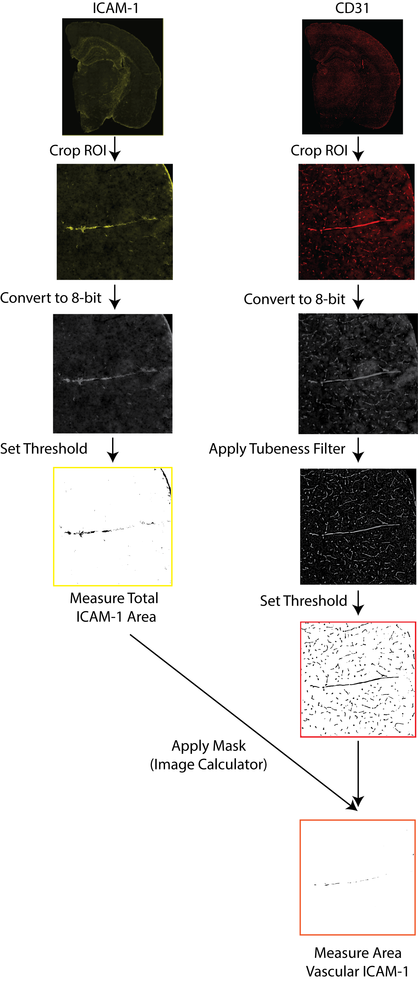
Fig. 2 Schematic diagram of quantification methods used for vascular immun‐
opositivity. Stained coronal brain sections were scanned in respective chan‐
nels, and ROIs were cropped and converted to 8-bit. The GFAP channel was
thresholded to remove background signals, while the ImageJ Tubeness filter
was applied to the CD31 channel to isolate the vasculature. Finally, the
filtered images were combined by the ImageCalculator function to select the
overlapping signals using the “AND” command.